With the development of science and technology, medical endoscopes have been widely used in the medical field. It is one of the important tools for humans to peek and treat internal organs. In the course of more than 200 years of development, the structure of endoscopes has undergone four major improvements, from the original rigid tube endoscope (1806-1932), semi-curved endoscope (1932-1957) to fiber endoscope endoscope (after 1957), and now the
electronic endoscope (after 1983). The image quality has also undergone a qualitative leap. Initially, the first rigid tube endoscope developed by the Germans used candlelight as the light source, and later changed to a light bulb as the light source. Nowadays, with LED lighting, the endoscope obtains color photos or color TV images. The image is no longer an ordinary image of tissues and organs, but a microscopic image observed under a microscope. Small lesions are clearly identifiable, and the image quality has reached a high level. Medical endoscopes are becoming more and more popular in clinical applications, and they are developing towards miniaturization, multi-function, and high image quality. The following introduces the classification, composition, structure, working principle, clinical application and development trends of medical endoscopes.
1. Classification of medical endoscopes
Classification according to its imaging structure: it can be roughly divided into three categories: rigid tube endoscopes, optical fiber endoscopes and electronic endoscopes. However, electronic endoscopes (can be divided into soft endoscopes and hard endoscopes)
Classified according to its function:
Endoscopes used for the digestive tract: rigid tube esophagoscope, fiber esophagoscope, electronic esophagoscope, ultrasonic electronic esophagoscope; fiber gastroscope, electronic gastroscope, ultrasonic electronic gastroscope; fiber duodenoscope, electronic duodenoscope Endoscope; fiber enteroscope, electronic enteroscope; fiber colonoscope, electronic colonoscope; fiber sigmoidoscope and proctoscope.
Endoscopes for the respiratory system: rigid laryngoscope, fiberoptic laryngoscope, electronic laryngoscope; fiberoptic bronchoscope, electronic bronchoscope;
Endoscopes used in the peritoneal cavity: rigid tube type, optical fiber type, electronic surgical laparoscope
Endoscopes used for the biliary tract: rigid choledochoscope, fiberoptic choledochoscope, and electronic choledochoscope.
Endoscopes used in the urinary system: Cystoscopes: can be divided into inspection cystoscopes, ureteral intubation cystoscopes, surgical cystoscopes, teaching cystoscopes, photography cystoscopes, pediatric cystoscopes and female cystoscopes . Ureteroscopy. Nephroscopy.
Endoscopes used in gynecology: hysteroscopy, abortion scope, etc.
Endoscope for joints: arthroscopy.
2. Composition of medical endoscope system
The medical endoscope system is generally composed of three major systems:
Looking glass system----Image display system-----Lighting system
Endoscope consists of: mirror.
The image display system consists of: CCD photoelectric sensor, display, computer, and image processing system.
The lighting system consists of: lighting source (xenon lamp cold light source, halogen lamp cold light source, LED light source) and transmission beam.
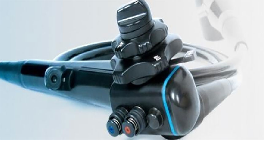
Fiber endoscopes generally consist of eyepieces, handwheels (soft or semi-hard), jaw openings, light guide interfaces, image guide beams, and light guides. Some products also include water (air) holes, obturators, etc. Fiber endoscopes are composed of optical observation systems, illumination transmission systems and bracket components. The optical observation system consists of an objective lens group for focusing imaging, a transmission/transmission group for transmitting the image of the objective lens group, and an eyepiece or CCD adapter lens for visual observation; the illumination transmission system is composed of multiple bundles of light guide fibers arranged in a mixed arrangement; the bracket The components are made of medical metal or organic materials that support and wrap the aforementioned system and have surgical or irrigation channels.
4. Classification of fiber endoscopes
Fiber endoscopes are divided into upper gastrointestinal tract endoscopes, lower gastrointestinal tract endoscopes and respiratory tract endoscopes according to their uses.
According to the optical viewing angle, it is divided into three types: front-view type, strabismus type, and side-view type.
According to function, it is divided into two types: with surgical function (with surgical and/or irrigation holes) and without surgical function (for examination).
5. Working principle of fiber endoscope
The biggest difference between fiber endoscopes and traditional endoscopes composed of pure optical lenses or electronic endoscopes is that the image transmission group uses image transmission optical fiber (optical fiber), which consists of multiple bundles of light guide fibers aligned according to coordinates In principle, in an area array arrangement, each light guide fiber serves as a pixel on the area array, corresponding to the coordinate positions at both ends of the image transmission fiber. The objective lens directly focuses and images the object on the fiber array. Each pixel (each light guide fiber) on the fiber array receives the light energy of the corresponding position image and transmits the light energy to the other part of the image transmission fiber. Emitted from one end, all the light energy output by all the pixels on the fiber array at the image side reorganizes the focused image of the objective lens, which achieves the purpose of fiber image transmission.
The key performance for the effective application of fiber endoscopes is the imaging level. In addition to requiring the objective lens to have a large viewing angle, small distortion, high relative aperture and depth of field, the quality of the image transmission fiber is the main contribution to the imaging quality and level of the fiber endoscope. Among them, the image transmission fiber The number of pixels in a fiber is a key factor that limits the resolution limit of a fiberscope (for a given field of view). The production of high-pixel digital imaging optical fiber involves single fiber core diameter manufacturing capabilities and molding technology. Such manufacturing processes include: hot melt method, etc. Arrangement processes include: single-layer lamination method, etc. At present, the minimum core diameter of image transmission optical fiber is less than 5 microns. Others, such as the consistency quality of single optical fiber and surface shape processing, also limit the quality of imaging optical fiber.
6. Introduction to image transmission optical fiber
Principle of optical fiber light transmission: Ordinary optical fiber is a cylindrical filament with a double-layer structure: a fiber core with a high refractive index in the middle, and a fiber coating with a low refractive index on the outside. There is good friction between the core and the coating. optical interface. Then the incident light will emerge from the other end of the optical fiber after several total reflections in the optical fiber. This is the simple principle of light propagation in optical fibers.
7. Composition of electronic endoscope
Electronic endoscopes are mainly composed of four main parts: endoscope, image processor, light source and monitor. Its imaging mainly relies on a miniature image sensor equipped on the front end of the lens body. The main function of the sensor is to convert light signals into electrical signals. It is a miniature camera that processes images through an image processor and then displays them on the monitor screen. The image is clearer, more realistic in color and higher resolution than ordinary optical fiber endoscopes, and can be viewed by multiple people at the same time.
8. Imaging principle of electronic endoscope:
The imaging principle of electronic endoscope is to use the light emitted by the light source equipped in the TV information center to guide the light into the body cavity under examination through the light guide fiber in the endoscope. The CCD image sensor receives the light reflected from the mucosal surface in the body cavity. This light is converted into electrical signals, and then the signals are transmitted to the television information center through wires. These electrical signals are then stored and processed through the television information center, and finally transmitted to the monitor to display the color of the examined organ on the screen. Mucosal images.
Advantages of electronic endoscopy in clinical applications:
Simple, flexible and convenient operation
Due to the application of electronic technology, when diagnosing and treating diseases, the operator, assistants and other staff can perform various operations under the direct view of the monitor, so that all operators can cooperate tacitly and safely. Therefore, it is flexible, convenient and easy to operate.
Patient discomfort is minimized
Due to the narrow diameter of the endoscope body, the patient's discomfort is minimized when the body is inserted into the body cavity.
Greatly improved diagnostic capabilities
Due to the application of CCD, the number of pixels is greatly increased compared with fiber endoscopes, the images are clearer and more realistic, and it has a magnification function. Therefore, it has a high resolving power, and it can observe the fine structure of the gastric mucosa, that is to say, it can observe the smallest anatomical unit of the gastric mucosa - gastric cells and gastric grooves. Therefore, tiny lesions can be found to achieve the ultimate goal of early detection, early diagnosis, and early treatment. In addition, due to the wide field of view of the electronic endoscope and the large bending angle of the front end of the endoscope, blind spots are avoided and missed diagnoses are avoided.
Convenient for teaching and clinical case discussion
Since the images are observed on the monitor screen, more people can observe and study together and conduct case discussions. At the same time, it also provides good conditions for improving the diagnostic level.
Facilitate close cooperation with patients
Since the images are observed on the monitor, the patient himself can also directly participate in the observation, which plays a positive role in eliminating the patient's nervousness and improving the patient's interest and confidence in the examination.
Provide reliable information for teaching and scientific research
Because electronic endoscopes can videotape and take photos of the inspection process, they can provide real and reliable first-hand information for future teaching and scientific research.
Development of electronic endoscope functions in clinical applications
Electronic endoscopy is the clinical endoscopy equipment with the most complete functions and the most development prospects following Kailai endoscopy. In addition to the application of electronic technology that makes images clearer and more realistic, the development potential of CCD alone is still quite huge. So far, CCD has reached 500,000 pixels, and according to data, it can reach up to 2 million pixels. It is conceivable that the electronic content In the process of continuous development and improvement of the mirror in the future, its image resolution will increase many times, and it will bring the detection of early lesions into a brand new world.
In addition, you can also use the TV information center to adjust red, blue, and green, and adjust different colors to observe different organizational structures, thereby achieving the best resolution of various organizational structures. At present, in addition to observing the smallest anatomical units of the gastric mucosa (gastric cells and minor grooves), electronic endoscopy can also observe the villous changes of intestinal metaplasia in the mucosa, regenerated epithelium around ulcers, new blood vessels, and submucosal Blood vessels and other tissue structures under the microscope, therefore, its ability to observe and diagnose diseases has reached a peak.
People apply image analysis technology to electronic endoscopy to obtain gastric blood flow maps, quantitative analysis of lesions, and temperature measurement. Ultrasound probes can also be installed on the front end of the endoscope for intracavity ultrasound exploration. In addition, communication lines can also be used to transmit electronic endoscopic images to distant locations for clinical disease consultation. In short, multi-functional electronic endoscopy will increasingly make great contributions in the diagnosis, treatment and study of disease pathogenesis and pathological changes in clinical diseases.
Rigid endoscope classification:
According to the viewing angle: (the angle between the viewing axis and the main axis of the mirror body)
1.3.1. 0° viewing angle rigid tube endoscope
1.3.2, 30° viewing angle rigid tube endoscope
1.3.5, 45° viewing angle rigid tube endoscope
1.3.3, 60° viewing angle rigid tube endoscope
1.3.4, 70° viewing angle rigid tube endoscope
1.3.5, 90° viewing angle rigid tube endoscope
10. Structure and composition of rigid tube endoscope
Rigid tube endoscopes are mainly composed of optical imaging systems and lighting systems.
The optical imaging system consists of three major systems: objective lens system, image rotation system, and eyepiece system.
Introduction to rigid tube endoscope objective lenses
Introduction to ordinary objective lenses: Ordinary objective lenses generally consist of 2---4 optical lenses to form the objective optical system.
. Its advantages are high resolution and good image quality. The disadvantages are complex structure, large size and high cost. The outer diameter of the lens can reach 1 mm in China and 0.6 mm in foreign countries.
Introduction to self-focusing lens objective lenses: also called variable refractive index lenses. It is a cylindrical lens made through special glass smelting, drawing, ion exchange, optical cold processing, and coating. The refractive index of the optical material of the lens changes according to a certain pattern along the radial direction. When light passes through, it produces the effect of converging light. Its advantages are cylindrical structure, simple shape, small size, the minimum diameter can reach 0.2 mm, high center resolution, especially suitable for making ultra-small endoscopes, low cost and easy assembly. The disadvantages are low edge resolution, poor uniformity and consistency.
11. Working principle of rigid endoscope
Working principle: The inverted image of the object under observation is formed by the objective lens. The inverted image is converted into an upright image through the image conversion system and transmitted to the eyepiece. It is then magnified by the eyepiece and observed by the human eye. In order to form different viewing angles, different prisms need to be added. Endoscopes for different purposes are made into different shapes, outer diameters, and lengths according to the requirements of use, so as to meet the requirements required for use.
The lighting transmission system consists of optical fibers. Working principle: The light from the cold light source is transmitted to the front end of the endoscope through the optical fiber to illuminate the object being observed.
As an important part of modern medicine, endoscopic technology provides doctors with deeper and more accurate diagnostic methods. Its continuous development and innovation will continue to promote the progress of medicine and bring better medical experience and treatment effects to patients.


