In the intricate world of histology, pathology, and biomedical research, precision is paramount. One of the most essential tools enabling such precision is the microtome. Despite being a staple in laboratories for decades, many people outside the scientific community are unfamiliar with this vital instrument. In this article, we will explore what a microtome is, its types, how it works, applications, maintenance, and future trends — providing a comprehensive understanding of this indispensable piece of equipment.
If you're looking for top-quality microtomes, be sure to explore our Microtome Collection at YSENMED, where professional solutions meet advanced technology.
What is a Microtome?
A microtome is a specialized instrument used to cut extremely thin slices of material, known as sections. These sections are typically examined under a microscope for biological or material science research. Microtomes allow scientists to produce sections thin enough for light to pass through, making cellular structures visible at high magnifications.
Microtome sections are critical for diagnosing diseases, conducting research, and analyzing the fine structures of tissues and materials. Precision, reproducibility, and consistency are the hallmarks of an excellent microtome, making it indispensable in laboratories worldwide.
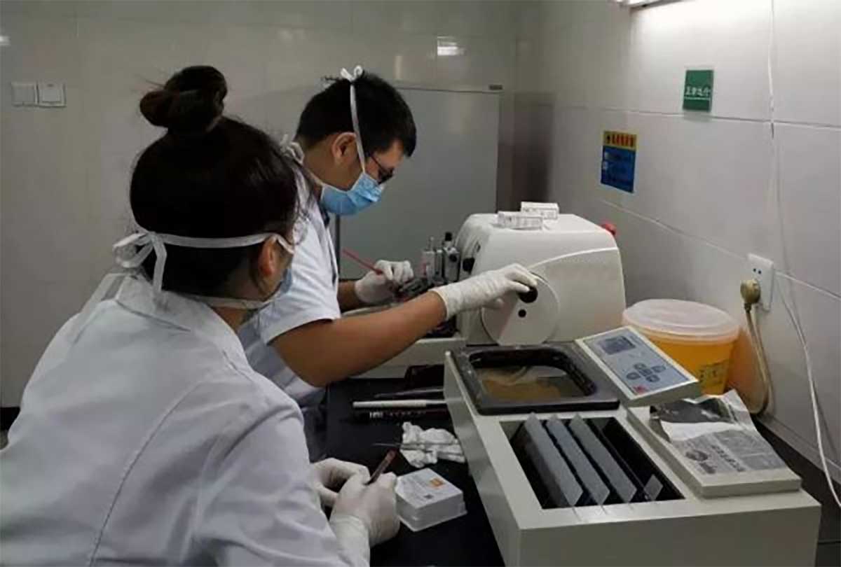
Types of Microtomes
There are several types of microtomes, each designed for specific applications:
-
Rotary Microtome: Most common type, ideal for routine histology. The specimen moves up and down while the blade remains stationary.
-
Sliding Microtome: Best for cutting hard tissues such as bone. Here, the knife moves horizontally across a stationary block.
-
Cryostat Microtome: Integrated with a freezing chamber, this microtome is used for cutting frozen sections for rapid diagnosis, especially in surgical settings.
-
Ultramicrotome: Designed for cutting ultra-thin sections required for electron microscopy.
-
Vibrating Microtome: Uses a vibrating blade to section delicate tissue without causing significant damage.
-
Sledge Microtome: Heavier and used for sectioning large or hard samples.
Each type of microtome offers unique advantages, making them suitable for different laboratory and research settings.
If you want to explore the best models available, check out our full range here: Microtome Collection.
How Does a Microtome Work?
The microtome operates by advancing a specimen against a very sharp blade to produce uniform sections. The process involves:
-
Specimen Preparation: Biological tissues are fixed, dehydrated, and embedded, usually in paraffin.
-
Setting Section Thickness: Adjust the micrometer to control slice thickness, typically from 1 to 100 microns.
-
Advancing the Block: The specimen is moved forward by the advancement mechanism.
-
Cutting the Section: Movement of either the block or blade slices the tissue into thin sections.
-
Section Collection: Carefully collected with brushes or floated in a warm water bath for mounting on slides.
-
Staining and Examination: After mounting, sections are stained and examined under a microscope.
Modern microtomes offer motorized advancements, precision control, and integration with imaging systems, significantly enhancing lab efficiency.
Step-by-Step Guide: Using a Microtome
To safely and effectively operate a microtome, follow these steps:
-
Preparation:
-
Loading the Specimen:
-
Setting Thickness:
-
Trimming:
-
Sectioning:
-
Collecting Sections:
-
Post-Processing:
-
Cleaning Up:
Safety and maintenance are critical when working with sharp instruments like a microtome.
Applications of Microtome
The microtome is indispensable across various scientific fields:
-
Medical Diagnostics: Pathologists rely on microtomes to diagnose cancers, infections, and tissue disorders.
-
Research: Researchers prepare samples for molecular studies, immunohistochemistry, and microscopic imaging.
-
Forensic Science: Analyzing tissues for legal investigations.
-
Botany: Examining plant structures and pathology.
-
Material Science: Cutting polymers and composite materials for structural analysis.
Without a microtome, detailed cellular studies and precise diagnoses would be significantly more challenging.

Choosing the Right Microtome: A Buyer’s Guide
Key factors when selecting a microtome include:
-
Sample Type: Soft tissue vs hard tissue dictates microtome type.
-
Section Thickness Requirements: Critical for imaging needs.
-
Level of Automation: Manual, semi-automatic, or fully automated options.
-
Safety Features: Finger guards, locking mechanisms, and emergency stop functions.
-
Durability: High-quality construction for long-term use.
-
Technical Support: Essential for training and maintenance.
At YSENMED Microtome Collection, we offer professional-grade microtomes engineered for precision and durability, suited for laboratories worldwide.
Maintenance and Care of Microtome
Maintaining your microtome is crucial for long-lasting accuracy:
-
Daily Cleaning: Remove wax and tissue debris after use.
-
Blade Care: Regularly replace or sharpen blades.
-
Lubrication: Keep moving parts well-lubricated.
-
Calibration: Frequently check the section thickness settings.
-
Annual Servicing: Partner with qualified technicians for inspections and servicing.
A well-maintained microtome ensures consistent, reliable results — protecting both the machine and user safety.
Future Trends in Microtome Technology
The microtome continues to evolve with advancements such as:
-
Smart Automation: Incorporating AI to enhance cutting precision and reduce human error.
-
Cryo-Microtomy Developments: Faster and more reliable frozen sectioning.
-
Eco-Friendly Materials: Sustainable manufacturing processes.
-
Integration with 3D Imaging: Enabling volumetric tissue studies.
Staying updated with these innovations ensures laboratories remain at the cutting edge of scientific discovery.
Frequently Asked Questions (FAQ)
Q1: What is a microtome used for?
A microtome is used for slicing extremely thin sections of tissues or materials for microscopic examination.
Q2: How thin can a microtome slice?
Depending on the model, microtomes can slice from 0.5 μm to several microns thick.
Q3: Is it difficult to operate a microtome?
With proper training and maintenance, using a microtome becomes straightforward and safe.
Q4: What materials can be sectioned using a microtome?
Tissues, plant material, polymers, and even composite materials can be sectioned.
Q5: Where can I buy a reliable microtome?
Visit YSENMED Microtome Collection for professional microtomes that meet all laboratory needs.
Conclusion
The microtome plays a pivotal role in science and medicine by enabling researchers and clinicians to explore the intricate details of tissues and materials. Its precision and versatility make it indispensable for histology, pathology, research, and beyond.
Whether you’re diagnosing disease, exploring cellular structures, or advancing scientific research, a high-quality microtome is an essential investment. Explore the comprehensive range of cutting-edge microtomes at YSENMED Microtome Collection and equip your lab for excellence today.
Choose precision. Choose reliability. Choose a microtome that advances your work toward the future.