1. How are X-rays produced?
The essence of
X-rays is electromagnetic waves with very short wavelengths, and the diagnostic wavelengths used in human medicine are 0.031-0.008nm. In the tube of the X-ray machine, the high-speed electron flow hits the metal target (tungsten, molybdenum). During the impact, the kinetic energy lost by the electrons’ sudden deceleration is released in the form of photons (the electrons transition from high energy level to low energy level and radiate electromagnetic waves), that is Can produce X-rays.
2. Composition of X-ray tube (ball tube)
X-ray tube is mainly composed of X-ray tube, X-ray machine power supply and control circuit.
Among them, the X-ray machine power supply is divided into high-voltage power supply and filament power supply. The function of the filament power supply is to heat the cathode filament; and the high-voltage output end of the high-voltage power supply is sandwiched between the cathode filament and the anode target, providing a high-voltage electric field to accelerate the active electrons on the filament to flow toward the anode target, forming a high-speed electron flow , to provide dynamic conditions for the electrons to strike the metal target.
The X-ray tube is the most important part of the X-ray generation process, mainly composed of a cathode filament, an anode target, and a vacuum glass tube. The cathode filament provides emitted electrons. The role of the anode target is to block the flow of high-speed electrons and impact them to produce Under current conditions, arc ignition occurs (arc is a gas discharge phenomenon, an instantaneous spark generated by current passing through certain insulating media), which damages the tube core and leads to tube scrapping or accidents.
Therefore, the process of X-ray generation in the emitter can be summarized as the filament heating generates a group of electrons that accelerate to the anode and hit the metal target under high voltage. After the electron flow bombards the anode target surface, 99% is converted into heat, and only 1% is due to X-rays are produced by strastrophtrophy (radiation produced by the sudden deceleration of high-speed electrons). Therefore, metal targets are generally large and rotating. Large-area and rotating metal targets are conducive to equipment heat dissipation and reduce equipment damage. This is also the reason why early X-ray machines were very large in size and noise.
3. X-ray characteristics and application imaging
Penetration and Absorption
The penetrability of X-rays refers to the fact that X-rays can pass through objects that visible light cannot pass through, and have different penetrating capabilities for materials of different densities. The penetrating capabilities are also related to the thickness and density of the material; the absorption of X-rays Sex refers to the attenuation of X-rays when penetrating materials. This is the basis of X-ray imaging. We can simply understand it as "advanced shadow".
photosensitive effect
X-rays can sensitize silver bromide (the photosensitive material in the film) to form a latent image. That is, the silver bromide (Ag+) in the film will turn into Ag (black) after being irradiated by X-rays, and the part blocked by the tissue will be There is no X-ray exposure or a small amount of exposure, resulting in a brighter "white" or "gray" image. The final X-ray image needs to be developed to appear, which is similar to the principle of early photo development.
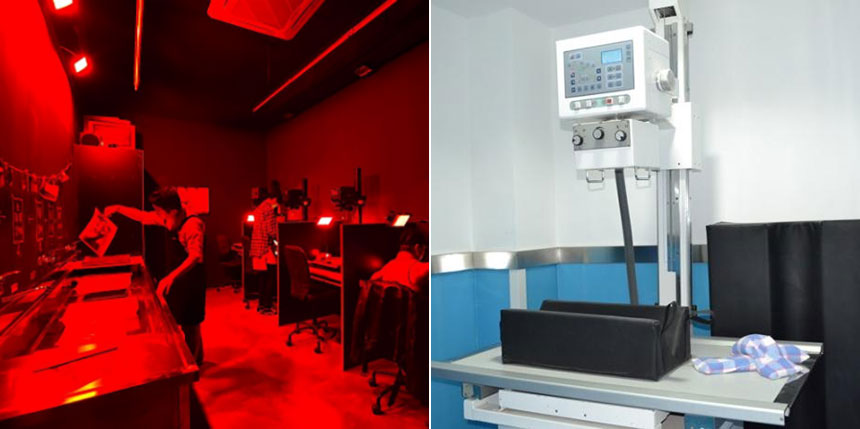
With the continuous advancement of technology, people use imaging boards (IP, DR boards) instead of film. After generating electrical signals through photosensitive elements, they are then processed by computers to form images. However, its essence is still a combination of the photosensitive effect of X-rays and technology. The old-fashioned X-ray machine used film to receive X-rays for imaging, while the new X-ray machines commonly used today use image plates to receive X-rays, so today's imaging doctors no longer have to go to the darkroom to "wash photos" in person.
fluorescent effect
X-rays can excite some fluorescent substances (calcium tungstate) to emit fluorescence (fluorescence is visible light). The most common fluorescence effect in life is to use the fluorescence effect of ultraviolet rays to distinguish genuine banknotes from counterfeit banknotes. This is the basic principle of X-ray fluoroscopy inspection . X-ray fluoroscopy was particularly popular in the 1980s and 1990s, and was mainly used for chest radiography examinations. Now it has been gradually replaced by other imaging equipment and out of use.
ionization effect
When X-rays pass through substances, they can ionize molecules and destroy molecular structures, which manifest in organisms as destroying proteins and nucleic acids. The biological effects produced by ionization are the basis of radiation protection and radiotherapy.
X-ray radiation has a cumulative effect, and long-term or frequent exposure to X-ray radiation has potentially serious harm to our bodies. X-ray radiation may affect or kill normal cells (especially immune cells) in animals. Medical research shows that high doses of electromagnetic radiation can also affect and destroy the original bioelectric current and biomagnetic field of the animal body; long-term exposure to high electromagnetic radiation environment will cause changes in blood, lymph and cell protoplasm, affecting the circulation of the animal body. Systemic, immune, reproductive and metabolic functions. At the same time, X-rays can also induce cancer and accelerate the proliferation of cancer cells in animals. In severe cases, they can also induce cancer.
Therefore, we must avoid frequent exposure to environments with X-ray radiation, carry out standardized protective work when necessary, and follow the arrangements of professional medical staff.
pet clinical
In pet clinical practice, radiation protection for Baoding assistants and pet owners should mainly be considered. Managers should mainly consider the following issues:
Utilization of radiation-proof building structures (thickness of room walls, closing of room doors during filming);
Reasonable use of auxiliary methods and tools for securing;
Baoding assistants wear protective clothing (lead gowns, lead gloves, neck scarves, lead goggles) and operate equipment in a standardized manner;
Radiation subsidies for Baoding assistants;
Full communication and standardized process before pet owners self-reserve;
The above are the basic principles of
X-ray imaging. I hope that the content of this issue can provide some help to all imaging learners and workers. YSENMED will also launch tweets on X-ray equipment related knowledge in the future, so stay tuned!


