In recent years, with the development of the medical industry, it is necessary to use different
X-ray equipment for diagnosis of different diseased parts of patients, especially the widespread use of CT machines and medical electron accelerators, etc., for the early diagnosis of patients, Targeted therapy provides very important help. According to statistics, 1.2 billion patients and examinees receive radiological diagnosis and treatment each year nationwide; therefore, carrying out radiation health monitoring and controlling radiation hazards can effectively reduce the radiation doses of radiation workers and examinees. There are mainly the following types of X-ray imaging equipment commonly used in medical institutions:
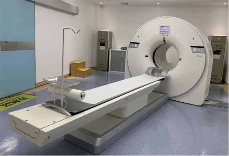
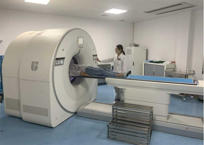
1. CT
CT (computerized tomography device) is a relatively advanced modern medical scanning inspection equipment. It can measure the human body with a highly sensitive instrument according to the difference in the absorption and transmittance of X-rays by different tissues of the human body, and then measure The obtained data is input into the electronic computer, and after the electronic computer processes the data, it can take a cross-sectional or three-dimensional image of the inspected part of the human body, and find small lesions in any part of the body. CT examination can examine all parts of the human body and discover the disease, with high resolution and sensitivity.
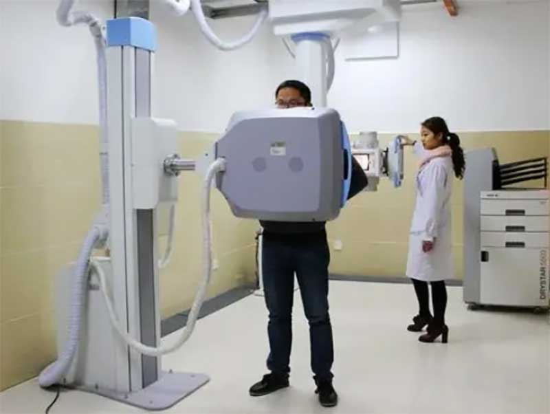
2. X-ray photography device
X-rays are emitted by the ball tube, and the rays pass through the detection object and reach the detector. According to the classification of detectors, they are divided into: DR, CR, and screen X-ray machines. Compared with CT images, the imaging is two-dimensional imaging.
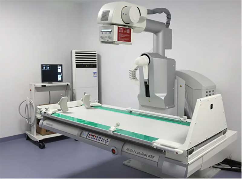
3. Gastrointestinal machine
Gastrointestinal machine is a device that integrates perspective and photography. For gastrointestinal fluoroscopy and photography, such as gastroenterography, esophagography, chest photography, myelography, arthrography, cholangiography, bronchography, venography, peripheral angiography, urography, hysterosalpingography, pediatric imaging , Partial interventional radiotherapy; it can also be used for skull and whole body bone photography, for fracture reconstruction and removal of foreign bodies under fluoroscopy.
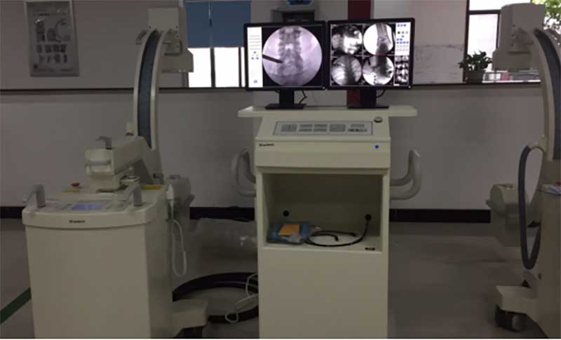
4. X-ray machine
Also known as X-ray fluoroscopy machine, portable X-ray fluoroscopy machine, it is an instrument that sees through objects and items through X-rays. X-ray fluoroscopy machines can penetrate many objects, such as glass, fiber, clothing, bone, blood, skin, tissue, wood, plastic, paper, thin metal and other media.
When X-rays pass through the human body, they are absorbed in different degrees. For example, the amount of X-rays absorbed by bones is more than that absorbed by muscles, so the amount of X-rays after passing through the human body is different, which carries information about the density distribution of various parts of the human body. , There is a big difference in the strength of the fluorescent effect or photosensitive effect caused on the fluorescent screen or on the photographic film, so shadows of different densities will be displayed on the fluorescent screen or on the photographic film (after developing and fixing). According to the comparison of shadow shades, combined with clinical manifestations, laboratory results and pathological diagnosis, it can be judged whether a certain part of the human body is normal. Such as orthopedic perspective, dental perspective and so on.
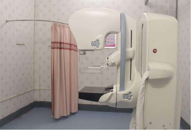
5. Breast machine
Mammography machine, also known as molybdenum target X-ray machine, is mainly used for X-ray examination of female breasts. It is the basic breast examination and diagnosis equipment in gynecology in hospitals. It can detect lumps and tiny tumors in breast tissue in time. It can also be used for photography of non-metallic foreign bodies and other soft tissues such as hemangioma.
Usually there are targets such as Mo/Rh/W. According to the needs of medical institutions for soft and hard rays, filters such as Mo/Rh/Al/Ag are usually configured. Detectable targets: Mo/Mo, Mo/Rh, Rh/Rh, W/Rh, W/Ag.
6. DSA
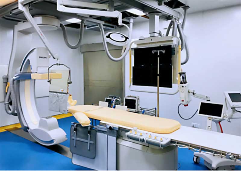
DSA (Digital Silhouette Angiography Equipment), with the development of imaging technology, more and more hospitals have begun to install such "tens of millions" of large-scale equipment. DSA has become essential for interventional surgery, especially for endovascular interventional technology. device of.
7. Mobile X-ray equipment
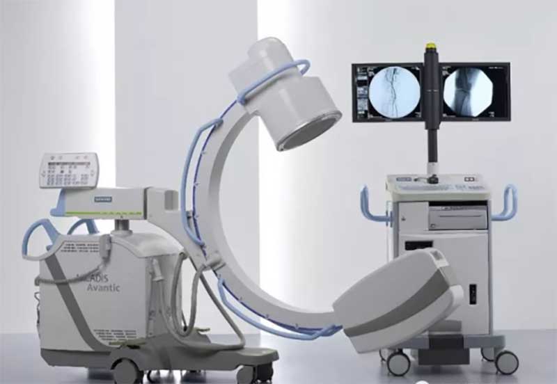
Mobile equipment has no fixed place of use. According to its imaging principle, it can be divided into mobile C-arm machine, mobile DR, mobile CR, and mobile screen X-ray machine (rarely used). It is suitable for radiology, orthopedics, wards, and emergency rooms. , operating room, ICU, etc., to meet the work needs of digital photography of the head, limbs, chest, spine, lumbar spine, abdomen and other parts of the body.
8. Crusher
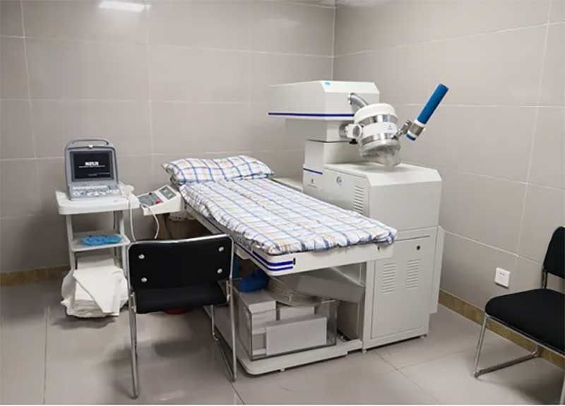
Extracorporeal shock wave lithotripsy (ESWL) is a shock wave generated by an extracorporeal lithotripsy machine, which is focused by the machine and aimed at the stones. The X-ray machine is used in conjunction with the X-ray system to locate the stones, and its properties are similar to those of the X-ray machine.
9. Molecular imaging equipment SPECT/CT and PET/CT
Single-Photon Emission Computed Tomography (SPECT) and Positron Emission Tomography (PET) are two imaging techniques in nuclear medicine, since they both detect gamma Radiography, also known as emission computed tomography (Emission Computed Tomography, ECT). The combined use of ECT and X-ray CT can more precisely locate the location, nature and extent of lesions.
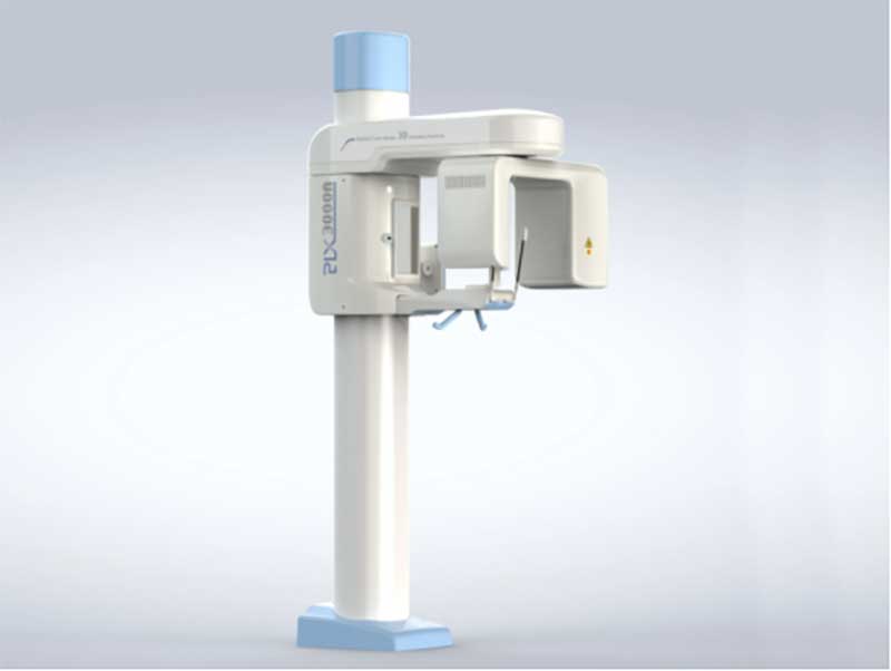
10. Dental X-ray machine
It is the general term for the intraoral dental film machine, panoramic machine and oral CT that we have seen so far, and it is the standard configuration of modern dentistry.
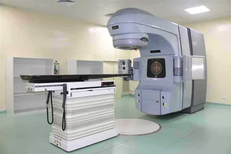
11. Linear accelerator
According to the types of accelerated particles, it can be divided into electron, proton and heavy ion linear accelerators. At present, electron accelerators are widely used, which can directly irradiate tumors in patients by generating X-rays and electron rays, so as to achieve elimination or reduction. The purpose of small tumors.
The above is to introduce the more commonly used X-ray equipment. If you are interested in X-ray equipment, you can contact us at any time. We are a professional manufacturer of
X-ray equipment.