1. Historical development
Since the German physicist Roentgen discovered X-rays on November 8, 1895, X-rays have been more and more widely used in medicine. In the early 20th century, a new discipline of radiology was born, and medical diagnosis and treatment began. In the new era of X-ray diagnostic technology, for more than a century, the rapid development and widespread popularization of X-ray diagnostic technology has made great contributions to human disease diagnosis and health care, and has always occupied the largest share in various branches of ionizing radiation medical applications. With the development of science and technology, the development of digital X-ray equipment is advancing by leaps and bounds, and has become the mainstream of medical X-ray diagnostic equipment. Radiography, digital mobile photography, digital gastrointestinal machine, etc.
2. Basic composition
X-ray generator
It refers to the device that completes the x-ray generation and controls it. It generally includes X-ray source components, high-voltage generators and control devices.
X-ray imaging device
X-ray imaging device: flat panel detector, computer system and image processing software, etc. The flat panel detector converts the X-ray signal into a digital signal and transmits it to the computer for processing. After the signal is processed by the computer, a digital X-ray film is obtained. After the digital film is processed by image software, each image can be automatically optimized according to different parts. It is to automatically adjust the gray scale of the over-bright and over-dark areas in the original exposure image, so that the brightness of each anatomical structure in an image is more uniform and coordinated, thereby improving the brightness of the over-bright or over-dark areas in the original exposure image. Ability to display contrast and detail. In addition, there are many other special software, such as automatic noise control, long limb stitching imaging, energy subtraction, automatic quality control, etc. The application of these software helps to make full use of the advantages and strengths of digital X-ray photography. One acquisition can Images with different effects and functions can be obtained to fully meet the needs of clinical diagnosis.
X-ray attachment
Refers to all kinds of supporting facilities designed to meet the needs of clinical diagnosis and treatment with X-ray generating devices. It mainly includes mechanical equipment such as diagnostic bed, support structure, suspension device, brake device, beam beam device, etc.
Digital radiography
Digital X-ray photography (DR) refers to a new technology of digital X-ray photography directly under the control of a computer, that is, the X-ray signal penetrating the human body is converted into a digital signal by an amorphous silicon (mainstream) flat panel detector, and the The computer reconstructs the image and performs a series of image post-processing. Compared with traditional optical imaging, it has the advantages of clearer images, lower radiation dose, faster inspection speed, and higher inspection success rate. DR imaging speed is fast, and radiographers can observe images on the screen within a few seconds. It can be sent to the post-processing workstation in a few seconds for image reading and diagnosis report, which greatly shortens the time from inspection to diagnosis report.
1: Composition of DR
The components of DR: Suspension system, ball tube, light beam device, photography bed, column, flat panel detector, exposure hand brake, shooting control system and image post-processing.
2: Imaging advantages
Amorphous silicon is directly converted into electrical signals after being irradiated by X-rays to obtain high-definition images. DR imaging has high spatial resolution, low noise rate, and high density resolution. DR photos can clearly see the mediastinum and lung tissue lesions behind the heart shadow by adjusting the window width, window level conversion curve and other technologies, and the ribs overlapping with the diaphragm can also be clearly displayed. Targeted image processing to improve the diagnostic rate.
3: DR application
At present, the areas where digital X-ray photography is used as the preferred or commonly used inspection method are roughly as follows:
Respiratory system
Digital X-ray photography is the basic examination method for the diagnosis of respiratory diseases, and it is also the most effective and economical method for screening. Because the living elbow tissue is filled with air and has natural high contrast, it is very suitable for X-ray photography examination. Chest X-ray plain film can provide an overview image of the chest, with high contrast and rich layers, and can detect most chest lesions. The disadvantage of X-ray chest plain film is that it is easy to miss the diagnosis of small lung lesions and overlapping lesions, and it is difficult to make a qualitative diagnosis of the lesions.
Musculoskeletal system
Due to the good natural contrast between bone and soft tissue and the simple and low-cost examination method, digital X-ray photography is still the preferred imaging examination method for bone and joint diseases. Plain x-ray films can not only show the extent and extent of lesions, but also make a qualitative diagnosis, but plain x-ray films cannot directly show soft tissue diseases such as muscles, tendons, menisci, and intervertebral discs, and it is not easy to find early-stage diseases of bone joints and soft tissues. lesion.
Abdomen
The application of ordinary X-ray photography in the abdomen is mainly for the diagnosis of acute abdomen, such as intestinal obstruction, gastrointestinal perforation, acute gastric dilatation, etc. Except for a few cases, conventional X-ray plain film can provide most of the diagnostic information.
Urinary System
Abdominal X-ray plain films can be used to display positive urinary stones. Intravenous nephrography can not only display the anatomical shape of the renal pelvis and ureter, but also judge the renal excretory function. It is a common imaging method for urinary system diseases.
Facial Features
The anatomical structure of the Wuxiao part is complicated and there are many fine structures. The display effect of digital X-ray photography on this part is not ideal, and it is difficult to meet the diagnostic requirements for most diseases. Instead, digital radiography is only used in the examination of a small number of diseases.
Eyes
X-ray plain film is mostly used for foreign body localization after eyeball trauma.
Mammography is a low-dose mammography technique. It can clearly display all layers of breast tissue, can find various benign and malignant breast lesions, and can observe tiny calcifications less than 0.1 mm; it is a highly effective and reliable inspection method for early detection and diagnosis of breast cancer, especially for clinically impossible and Early breast cancer with microcalcification as the only manifestation. The digital mammography system has the characteristics of clear imaging, convenient operation, fast inspection, and low radiation dose. It is an important inspection method for breast diseases and the "gold standard" for breast cancer screening recommended by the US FDA.
1: Development history
1913
German surgeon Salamon first used X-ray photography in the examination of breast diseases.
1965
Mammography tubes are used for mammography.
1973
The Frenchman Gros applied the rotating anode mammography X-ray tube to the mammary X-ray machine, and the automatic exposure control (AEC) and the compressor were used on the mammogram machine in the same year.
1976
Grids are used in mammography.
1981
0.1mm X-ray tube enabled.
1996
Charge-coupled devices (CCDs) are used in mammography machines.
2000
Emergence of fully digital mammography (FFDM)
year 2002
Computer-aided detection (CAD) is used for breast imaging.
year 2006
Clinical application of digital breast tomography (DBT).
year 2011
Enhanced mammography is used clinically.
2: Basic structure
The components of mammography: high voltage generator, X-ray tube imaging system, shooting control system, mechanical part.
3: How much do you know about mammography?
Are mammograms harmful to humans?
The radiation dose of routine mammography is low and will not endanger women's health, but normal women do not need to undergo repeated mammography in a short period of time.
Who needs a mammogram?
Women over the age of 40 get mammograms every year, and women between the ages of 30 and 40 get a mammogram at least every three years.
Who may be at high risk of breast cancer and need regular mammography?
• Have a history of breast cancer themselves.
• Family history of breast cancer: Family history of breast cancer in first-degree relatives, especially in bilateral or premenopausal relatives.
• Past history of the following breast diseases: such as ductal epithelial dysplasia, lobular neoplastic lesions (atypical lobular hyperplasia and lobular carcinoma in situ), juvenile papillomatosis, etc.
• Previous history of high-dose radiation therapy to the chest, such as radiation therapy for lymphoma.
• Early menarche and early establishment of regular menstrual cycles.
• Late age at menopause.
• Late age of first pregnancy (>30 years old) with only one child and no breastfeeding.
• Those who have never had children.
• Being overweight or obese before or after menopause.
• Hormone replacement therapy.
Digital gastrointestinal machine
Digital gastrointestinal machine is mainly used to check the X-ray examination equipment for gastrointestinal diseases. It can perform throat, esophagus, stomach, duodenum, jejunum and colon, urography, gynecological photography (hysterosalpingography), etc. Multi-faceted inspection is one of the main inspection methods for various ulcers, tumors, foreign bodies and other diseases. At the same time, the digital gastrointestinal tract can also perform fluoroscopy, and some vascular and non-vascular interventional treatments.
1: Composition of digital gastrointestinal machine
The digital gastrointestinal machine is mainly composed of high heat capacity tube, large-size dynamic flat panel detector, high-power high-voltage generator, beam beam device, multi-functional examination bed, high-speed perspective acquisition control system, image post-processing software and other parts.
2: Features of digital gastrointestinal machine
• The image quality is good, and the image is collected digitally. All the collected images are digitally processed and the images are clearer, so that doctors can observe subtle lesions under various conditions, thereby improving the accuracy rate.
• Timely dynamic image acquisition can adjust the image acquisition speed according to different inspection parts, and can observe whether the physiological movement of organs changes.
• The timely function of digital subtraction angiography can cut some overlapped images in conventional angiography, so as to improve the image quality and reduce the rate of misdiagnosis and missed diagnosis.
• The fluoroscopy dose is significantly reduced, greatly reducing the adverse effects of radiation on patients and staff.
DR is the abbreviation of digital direct imaging system. Compared with traditional X-ray imaging, it has the advantages of clearer image, lower radiation dose, faster inspection speed and higher inspection success rate. Bedside digital mobile DR photography technology emerged as the times require, and is suitable for radiology, orthopedics, wards, emergency rooms, operating room ICU and other departments to meet the needs of mobile photography such as chest, limbs, abdomen, and bedside positioning.
1: Structure of Mobile DR
The mobile DR machine is mainly composed of a high-voltage generator, a ball tube, a flat panel detector, a light beam device, an acquisition control system, an image processing workstation, and a mechanical device.
2: The working process of mobile DR
X-rays pass through the human body (parts for inspection) and are projected onto the flat-panel detector, and then the detector directly converts the X-ray signal into a digital signal and transmits it to the image workstation synchronously, and the image is post-processed by the professional medical software of the workstation to obtain A digital radiograph.
3: Features of Mobile DR
The mobile DR machine is different from the column-type or suspension-type design of the traditional photography system. The mobile DR machine occupies a smaller space and is easy to move and operate. It can directly complete the exposure in the ward and supports multiple bedside imaging examinations. It not only effectively improves work efficiency , to meet the production demand, optimize the hospital workflow, and at the same time ensure the imaging quality, it is an indispensable equipment for radiological diagnosis.
The mobile DR machine can quickly acquire and confirm photographic images after a few seconds of exposure, eliminating the need for traditional film processing and IP board information reading and other complicated procedures. Images can be processed on site, network transmission, and printing, which is efficient and fast.
The basic principle of the digital angiography subtraction angiography system is to digitally input two frames of X-ray images taken before and after injection of contrast agent into the image computer, and obtain a clear pure vascular image through the process of subtraction, enhancement and re-imaging, and at the same time visualize the blood vessels in real time DSA has the advantages of high contrast resolution, short examination time, less contrast agent consumption, and low radiation dose. It is of great significance in the clinical diagnosis of vascular diseases. DSA is mainly used for observing vascular lesions, positioning measurement of vascular stenosis, and providing real three-dimensional images for interventional treatment, which is a necessary condition for various interventional treatments. It is suitable for the inspection and interventional minimally invasive treatment of cardiovascular and cerebrovascular, peripheral blood vessels and tumors.
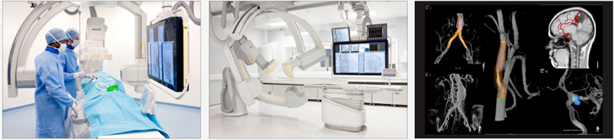
1: Clinical application of DSA
DSA is helpful for straightening of the heart and great vessels, and can be used for aortic dissection, aortic aneurysm, coarctation of the aorta or abnormal development of the aorta and for checking the pulmonary artery.
DSA is the gold standard for displaying coronary arteries.
It can clearly show the cervical and intracranial arteries, and can be used to diagnose cervical artery stenosis or occlusion, intracranial aneurysms, abnormal vascular development and arterial occlusion, as well as blood supply arteries and tumor staining of intracranial and intracranial tumors.
DSA can well display the abdominal aorta, its large branches, and extremity blood vessels.
2: New technology and prospect of DSA
In recent years, the macroscopic development trend of DSA is to change to specialization, that is, the one-way C-arm system is used for whole-body angiography and interventional radiology, and the two-way C-arm system is used for heart and cerebrovascular examination. Currently, the rotating DSA imaging The equipment has been used clinically, which can make the X-ray tube rotate or move with multiple tracks, and can realize the subtraction image display of three-dimensional angiography. At the same time, the method of storing masks in the computer or the realization of programmed step-by-step DSA has improved the Insufficiency of conventional DSA in the past, effectively improving the spatial resolution and time resolution of DSA images, reducing X-ray dose, further enhancing the inspection efficiency of the system, and improving the quantitative analysis ability of images are the main development trends of DSA, and also the main development trend of medical imaging diagnosis. The key to treatment.


